
Surgical Procedures
Expert Care. Proven Results. Trusted Hands.
Our practice specializes in advanced surgical dermatology, offering expert care in the diagnosis and treatment of skin cancer and other complex skin growths. Whether you’ve been referred for a biopsy or need skin cancer removal, our team is dedicated to providing the highest levels of safety, precision, and compassionate care every step of the way.
We evaluate, biopsy, and treat all forms of skin cancer, with a focus on early detection and evidence-based care. Every skin cancer removal we perform is pathology-confirmed to ensure accuracy and complete clearance.
Our Surgical Treatment Options
Request Appointment
Have a general dermatology concern? Schedule Today!
Call Rochester Hills
(248) 852-1900
Call Livonia
(734) 307-2400
Call Macomb
(586) 368-3600
Call Novi
(248) 946-4787
Call Allen Park
(313) 346-9900
Book Online
Common Skin Cancers We Treat
Skin cancer is the most common form of cancer in the United States, with over 5 million cases diagnosed annually. While this statistic may seem alarming, the vast majority of skin cancers are highly treatable when detected early. At Beacon Skin & Surgeries, we specialize in the comprehensive diagnosis and treatment of all types of skin cancer, from the most common basal cell carcinomas to the more serious melanomas. Our experienced team uses advanced diagnostic techniques and proven treatment methods to ensure the best possible outcomes for our patients, while our commitment to thorough follow-up care provides peace of mind throughout your treatment journey.
Basal Cell Carcinoma (BCC)
Basal cell carcinoma is the most frequently diagnosed form of skin cancer, accounting for approximately 80% of all skin cancer cases. This type of cancer develops in the basal cells, which are found in the deepest layer of the epidermis. BCC typically occurs in areas of the body that receive significant sun exposure, such as the face, neck, ears, scalp, shoulders, and back.
BCC often presents as a pearly or waxy bump, though it can also appear as a flat, flesh-colored or brown scar-like lesion, or a pink patch of skin. The lesion may have visible blood vessels, a central depression, or may bleed easily when touched. While basal cell carcinomas grow slowly and rarely metastasize (spread to other parts of the body), they can cause significant local tissue damage if left untreated. The cancer can grow deeper into the skin and underlying tissues, potentially affecting muscles, nerves, and even bone in advanced cases.
Treatment for BCC depends on the size, location, and subtype of the tumor. Options include surgical excision, Mohs micrographic surgery for high-risk or recurrent tumors, electrodesiccation and curettage for smaller lesions, cryotherapy, topical medications for superficial types, and radiation therapy in select cases. The prognosis for BCC is excellent when detected and treated early, with cure rates exceeding 95% for most treatment methods.
Squamous Cell Carcinoma (SCC)
Squamous cell carcinoma is the second most common type of skin cancer, developing in the squamous cells that make up the middle and outer layers of the skin. SCC can occur anywhere on the body but is most frequently found on sun-exposed areas such as the face, ears, neck, lips, and backs of the hands. This type of cancer can also develop in areas of chronic inflammation, previous burns, or scars.
SCC may present in various forms, including a persistent scaly red patch with irregular borders that may crust or bleed, an elevated growth with a central depression that occasionally bleeds, a wart-like growth, or an open sore that doesn’t heal or heals and returns. Unlike basal cell carcinoma, squamous cell carcinoma has a higher potential for metastasis, particularly when it develops in certain high-risk locations such as the lips, ears, or genitals, or when it arises in immunocompromised patients or areas of chronic inflammation.
Early detection and treatment of SCC are crucial because this cancer can grow deeper into the skin and spread to lymph nodes and other organs if left untreated. Treatment options include surgical excision with clear margins, Mohs micrographic surgery for high-risk tumors, electrodesiccation and curettage for smaller, low-risk lesions, radiation therapy, and in advanced cases, systemic therapies. The five-year survival rate for localized SCC is nearly 100%, but this drops significantly if the cancer has spread, emphasizing the importance of early intervention.
Melanoma
Melanoma is the most serious and potentially deadly form of skin cancer, developing in the melanocytes—the cells that produce melanin, the pigment that gives skin its color. Although melanoma accounts for only about 1% of all skin cancers, it causes the majority of skin cancer deaths due to its aggressive nature and tendency to metastasize rapidly. Melanoma can develop anywhere on the body, including areas not typically exposed to sun, such as the soles of the feet, palms of the hands, fingernail beds, and mucous membranes.
Melanoma often appears as a new mole or as changes in an existing mole. The ABCDE criteria help identify suspicious lesions: Asymmetry (one half doesn’t match the other), Border irregularity (ragged or notched edges), Color variation (multiple colors or uneven color distribution), Diameter larger than 6mm (size of a pencil eraser), and Evolving (changes in size, shape, color, or symptoms). However, melanomas can also present as amelanotic (non-pigmented) lesions that appear pink or flesh-colored, making professional evaluation essential for any changing or concerning skin lesion.
The treatment and prognosis of melanoma depend heavily on early detection and the depth of invasion (Breslow thickness) at the time of diagnosis. Treatment typically begins with wide surgical excision to remove the primary tumor with appropriate margins. Depending on the thickness and characteristics of the melanoma, additional procedures may include sentinel lymph node biopsy to determine if the cancer has spread to nearby lymph nodes. For advanced melanomas, treatment may involve immunotherapy, targeted therapy, chemotherapy, or radiation therapy. When caught early and confined to the skin surface, melanoma has an excellent prognosis with five-year survival rates exceeding 99%. However, once it spreads to lymph nodes or distant organs, the prognosis becomes more serious, underscoring the critical importance of regular skin examinations and immediate evaluation of any suspicious lesions.
Mohs Surgery
Mohs micrographic surgery is a highly precise surgical technique that removes skin cancer one microscopic layer at a time while preserving as much healthy tissue as possible. This advanced procedure is most commonly used to treat basal cell carcinomas and squamous cell carcinomas, particularly in cosmetically sensitive areas or recurrent cases.
What makes Mohs unique is that the surgeon examines each removed tissue layer under a microscope immediately after removal, ensuring complete cancer removal while minimizing healthy tissue loss.
Why Choose Mohs Surgery?
Mohs surgery offers several significant advantages over traditional skin cancer removal methods:

Highest Cure Rate
- Up to 99% cure rate for primary basal cell and squamous cell carcinomas
- 95% cure rate for recurrent skin cancers
- Superior outcomes compared to standard excision methods

Tissue Preservation
- Minimally invasive technique that removes only cancerous tissue
- Preserves maximum amount of healthy surrounding skin
- Ideal for areas where tissue conservation is critical (face, hands, genitals)

Same-Day Results
- Immediate confirmation that all cancer has been completely removed
- Real-time microscopic examination eliminates guesswork
- No waiting for pathology results or additional procedures

Outpatient Procedure
- Performed under local anesthesia in a comfortable office setting
- Most patients return to normal activities within a few days
- No hospital stay required
Our Mohs Expert
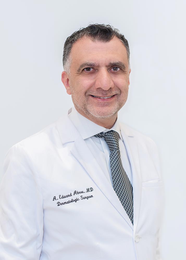
Dr. Abrou is a board-certified dermatologist with specialized fellowship training in Mohs surgery and years of dedicated surgical experience.
Advanced Capabilities
- On-site laboratory for same-day tissue processing and diagnosis
- State-of-the-art microscopy and surgical equipment
- Expert cosmetic repair and reconstruction by Dr. Abrou
- Same-day wound closure in most cases
What to Expect During Mohs Surgery
The Process:
- Local Anesthesia – The area is thoroughly numbed for your comfort
- Precise Removal – A thin layer of tissue is removed and carefully mapped
- Immediate Analysis – Dr. Abrou examines 100% of the tissue margin under microscopy
- Layer-by-Layer Precision – If cancer remains, additional tissue is removed only from affected areas
- Same-Day Repair – Once clear margins are achieved, the wound is repaired using the most appropriate technique for optimal results
The procedure typically takes 2-4 hours depending on the cancer’s size and depth. Patients have immediate confirmation that their cancer has been completely removed with the highest possible cure rate.
Before & After Mohs Surgery Examples

Surgical Excision
Not every skin cancer requires Mohs surgery. Standard surgical excision is often the best treatment choice for certain types of skin cancers, particularly melanomas and tumors located in low-risk areas where tissue preservation is less critical. This proven technique is also ideal for skin cancers with well-defined borders, smaller lesions, and cases where immediate pathology analysis isn’t necessary.
Surgical excision offers an excellent balance of effectiveness, efficiency, and cosmetic outcomes for appropriately selected cases.
Our Surgical Approach
Precise Technique with Cosmetic Focus
- Performed under local anesthesia for patient comfort
- Strategic incision planning to follow natural skin lines when possible
- Removal of cancer with appropriate safety margins based on tumor type and size
Comprehensive Pathology Analysis
- All tissue specimens sent to certified pathologists for thorough examination
- Detailed margin analysis to confirm complete cancer removal
- Additional treatment recommendations if margins are close or positive

Expert Wound Closure
- Closure techniques tailored to minimize scarring and optimize healing
- Consideration of skin tension, location, and individual healing factors
- Advanced suturing methods for the best possible cosmetic outcome
What To Expect
During the Procedure:
- Local anesthesia ensures comfort throughout the process
- Quick, efficient removal typically completed in 15-30 minutes
- Immediate wound closure with careful attention to cosmetic results
After Surgery:
- Clear, detailed post-operative care instructions
- Scheduled follow-up appointments to monitor healing
- Pathology results typically available within 5-7 business days
- Additional treatment planning if needed based on pathology findings
Recovery and Follow-up:
- Most patients experience minimal discomfort and rapid healing
- Activity restrictions provided based on location and size of excision
- Long-term follow-up care for ongoing skin cancer prevention
Biopsies
If your provider has recommended a skin biopsy, rest assured that it’s a quick, straightforward procedure performed safely in our office. A biopsy is simply the removal of a small tissue sample for microscopic examination, allowing us to make an accurate diagnosis and develop the most appropriate treatment plan.
Skin biopsies are one of the most valuable diagnostic tools in dermatology, providing definitive answers when visual examination alone isn’t sufficient to determine the nature of a skin lesion.
What We Evaluate Through Biopsy

Suspicious or Changing Lesions
- Changing or irregular moles that have altered in size, shape, or color
- New spots that have appeared and don’t look like your other moles
- Non-healing sores or spots that persist beyond normal healing time
- Lesions with unusual colors, borders, or surface characteristics

Growths and Nodules
- Suspicious bumps or nodules that appear concerning
- Cysts, lipomas, or other growths that are enlarging or causing discomfort
- Unusual skin textures or thickened areas
- Any lesion that doesn’t respond to standard treatments
The Biopsy Process
Simple Office Procedure:
- Performed under local anesthesia for maximum comfort
- Takes only 10-15 minutes depending on the size and location
- Minimal discomfort during and after the procedure
- Most patients return to normal activities immediately
Types of Biopsies:
- Shave biopsy for raised lesions or surface abnormalities
- Punch biopsy for deeper tissue sampling when needed
- Excisional biopsy for complete removal of smaller lesions
Expert Pathology Analysis:
- All tissue samples sent to trusted, board-certified dermatopathology laboratories
- Specialized pathologists with expertise in skin conditions
- Results typically available within 7–10 business days
- Detailed pathology reports provide comprehensive diagnostic information
After Your Biopsy

Comprehensive Follow-up Care
If further treatment is needed based on your biopsy results, we’ll guide you through every step of the process. Our team takes time to explain your diagnosis in understandable terms, discuss all available treatment options, and answer any questions you may have.

Patient Support and Education
- Clear explanation of biopsy results and their implications
- Detailed treatment recommendations tailored to your specific diagnosis
- Timeline for any additional procedures or follow-up care needed
- Ongoing support to ensure you feel informed and confident in your care
Our goal is to make the biopsy process as comfortable as possible while providing you with accurate diagnostic information and comprehensive follow-up care when needed.
FAQs
What To Expect For Surgical Visits
Not every skin cancer requires Mohs surgery. This proven technique is also ideal for skin cancers with well-defined borders, smaller lesions, and cases where immediate pathology analysis isn’t necessary.
Surgical excision offers an excellent balance of effectiveness, efficiency, and cosmetic outcomes for appropriately selected cases.
Mohs Surgery Before & After
Click to View: Back of Neck & Neck | Bridge & Nose | Cheek | Chin | Ear | Elbow | Eye & Eyebrow | Forehead, Head, & Temple | Hand & Wrist | Lip | Shin
Back of Neck & Neck

Before

During

After

Healed

Before

During

After

Healed

Before

During

After

Healed

Before

During

After

Healed
Bridge & Nose

Before

During

After

Healed

Before

During

After

Healed

Before

During

After

Healed

Before

During

After

Healed

Before

During

After

Healed

Before

During

After

Healed

Before

During

After

Healed

Before

During

After

Healed

Before

During

After

Healed
Cheek

Before

During

After

Healed

Before

During

After

Healed

Before

During

After

Healed

Before

During

After

Healed

Before

During

After

Healed
Chin

Before

During

After

Healed

Before

During

After

Healed
Ear

Before

During

After

Healed

Before

During

After

Healed

Before

During

After

Healed

Before

During

After

Healed

Before

During

After

Healed
Elbow

Before

During

After

Healed
Eye & Eyebrow

Before

During

After

Healed

Before

During

After

Healed

Before

During

After

Healed

Before

During

After

Healed
Forehead, Head, & Temple

Before

During

After

Healed

Before

During

After

Healed

Before

During

After

Healed

Before

During

After

Healed

Before

During

After

Healed

Before

During

After

Healed

Before

During

After

Healed

Before

During

After

Healed

Before

During

After

Healed
Hand & Wrist

Before

During

After

Healed

Before

During

After

Healed

Before

During

After

Healed

Before

During

After

Healed
Lip

Before

During

After

Healed

Before

During

After

Healed

Before

During

After

Healed

Before

During

After

Healed
Shin

Before

During

After

Healed
At Beacon Skin & Surgeries, we believe in transparency and education. The following images reflect real Mohs surgical procedures and may be graphic to some viewers. They are shared to provide a clear understanding of the care we deliver.